Should primary microsurgical ligation of varicocele be the gold standard approach?
Abstract
Introduction: Varicocele is a common condition affecting over one in 10 men, and in cases with abnormal semen parameters, varicocele is present in about one in four men. Several methods have been used to treat this condition, of which microsurgical treatment has the lowest failure and complication rates. We present a single-centre UK series of microsurgical repair of varicocele.
Methods: All microsurgical varicocelectomy cases performed between 2013 and 2017 were retrospectively identified and reviewed. The approach used is low inguinal using a high-powered Leica microscope.
Results: Microsurgical varicocele repairs were performed in 30 cases, with a median age of 34.7 years. There were 22 cases with chronic scrotal pain of which eight cases had failed previous embolisation with a success rate of 90.5%. There were eight with oligoasthenoteratozoospermia, showing an improvement of more than 75% in sperm count after repair.
Conclusions: Microsurgical ligation is a very effective treatment for symptomatic varicoceles and should be considered when radiological embolisation has failed. It is also effective in improving semen parameters in all followed up oligoasthenoteratozoospermia cases. It should be considered a primary method of varicocele treatment in the UK.
Level of evidence: 3.
Introduction
Varicocele is a common condition affecting over one in 10 men, and in cases with abnormal semen parameters varicocele is present in about one in four men (1). The treatment of varicocele is controversial but is recommended in subfertility as improvements in semen parameters are consistently observed after repair (2) as well as reversal in sperm DNA dam- age (3). Varicocele repair is also recommended in testicular underdevelopment and in symptomatic varicoceles (4). Several methods have been used to treat this condition; surgical open and laparoscopic as well as angiographic sclerotherapy and embolisation (5). Microsurgical treatment has the lowest failure rates compared to all treatment techniques including radio- logical embolisation; however, this requires microsurgical training, limiting its widespread use (5,6). We present a single- centre UK series of microsurgical repair of varicocele.
Methods
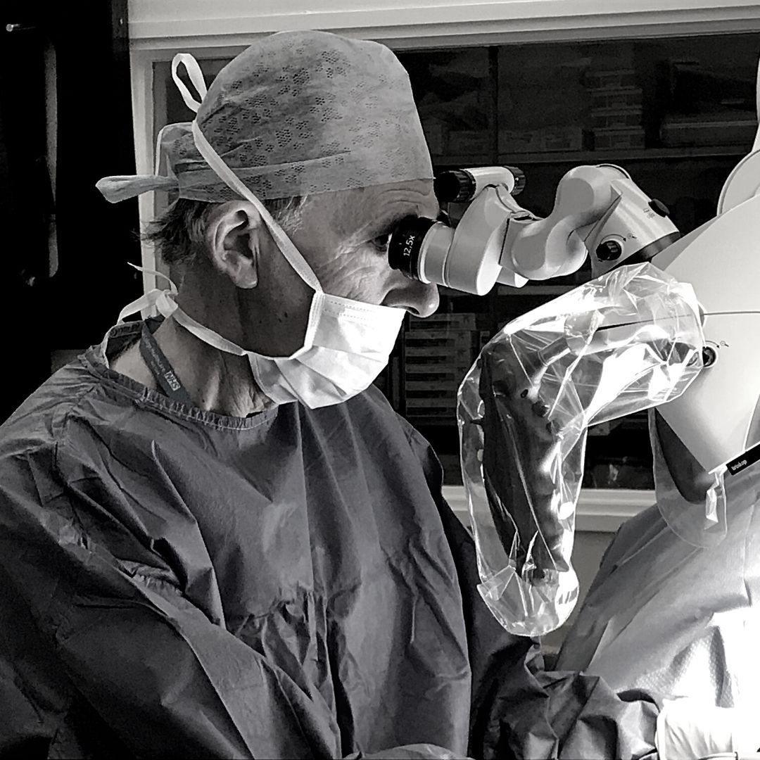
All microsurgical varicocelectomy cases performed between 2013 and 2017 were retrospectively identified and reviewed. The approach used was low inguinal inci- sion over the external inguinal ring. The cord and testicle are then delivered. Using a high-powered Leica microscope, the artery and lymphatics are identified and spared, and then all dilated veins are ligated, including accessory or gubernacular veins. Routine follow-up was organised at 6 and 12 weeks after surgery. In the chronic scrotal pain cohort, the treatment was con- sidered successful when pain has completely resolved. Semen analysis was repeated at the 12-week postoperative visit for oligoasthenoteratozoospermia patients and com- pared to the pre-treatment parameters.
Results
Between 2013 and 2017, microsurgical varicocele repairs were performed in 30 cases, with a median age of 34.7 years (range 18.7–68.2 years). The indications are summarised in Table 1.
Overall, there were 21 cases with chronic scrotal pain, of which 13 had microsurgical repair as the primary treat- ment while the remaining eight cases had failed previous radiological embolisation, of which three cases had two radiological embolisation treatments.
In the primary treatment group, 12 out of 13 (92.3%) had resolution of the pain on follow-up. While in the group that had previous radiological embolisation treatment, seven out of eight (87.5%) had successful resolution of symptoms following microsurgical repair, and that includes the three cases that had embolisation attempts twice.
Microsurgical ligation was performed for oligoasthe- noteratozoospermia in eight cases. Pre-treatment semen analysis showed a median sperm count of 5.7×10⁶ (range 7 sperms to 9×10⁶/ml), the forward motility median 31.5% (range 21–46%) and the normal morphology median 1.8% (range 0–2%). Post-treatment semen analy- sis was available in seven cases and they all showed sig- nificant improvement in the total semen count of over 75% (median 17.6×10⁶; range 1–33×106/ml; P=0.0274). Also the motility (median 40.5%; range25–49%) and morphology parameters improved (median 2.5%; range 1–4%). At least three of the oligoasthenoteratozoospermia cases reported subsequent successful natural pregnancy and childbirth.
There was one case of testicular underdevelopment, and the testicular size improved over a 3-year follow-up.
Postoperatively eight had postoperative bruising or small haematoma treated conservatively; one had sympto- matic cord haematoma needing release.
Discussion
The treatment of varcicocele has been highly debated in the literature. In the case of male infertility, initial reviews were not conclusive (7) and the National Institute for Health and Care Excellence (NICE) did not recommend offering varicocele treatment to subfertile men to improve fertil- ity (8). A 2012 Cochrane review including more randomised control trials9 showed evidence in favour of clinical varicocele repair in the case of subfertility and that it improves the couple’s chance of pregnancy (odds ratio 1.47, 95% confidence interval 1.05–2.05). Baazeem et al. revealed that varicocele repair enhances semen analysis parameters and reduces seminal oxidative stress and sperm DNA damage (4).
In our series, all patients with subfertility had primary microsurgical repair of their clinical varicocele. Patients who provided post-surgery semen analysis showed significant improvement in sperm parameters and an over 75% increase in total sperm count (P=0.0274), shifting the total count closer to or above the normal range.10 Also, three out of eight cases reported successful natural preg- nancy and childbirth.
According to the European Association of Urology guidelines, scrotal pain is one of the recommended indications to repair varicoceles (6) and they also discuss the several procedures by which this can be performed. Interventional radiological sclerotherapy and embolisation are less inva- sive methods that are used by many centres in the UK. This is associated with a small risk of thrombophlebitis, bleed- ing and reaction to contrast medium, as well as a reported failure rate up to 10%;11 this can be due to extensive venous branching or accessory or gubernacular veins. In our series, eight cases had previous failed embolisation and three had two failed embolisation procedures. Seven cases, including the three cases with two failed embolisations, were treated successfully with microsurgical repair. It was not possible to assess and directly compare the failure rate of radiologi- cal embolisation at our institution as most of these emboli- sations were done in other hospitals.
The downsides of a surgical approach are the risks of general anaesthesia, wound-related complications, delayed recovery time and return to work, risk of testicular arterial injury and subsequent testicular atrophy, lymphatic injury and subsequent hydrocele.
With the use of the microsurgical technique that was first described by Goldstein et al.12 in 1992, arterial and lymphatic vessels are recognised and spared while exten- sive venous branches can be identified easily and ligated. Also, accessory and gubernacular vessels, which may be the cause of failed radiological embolisation, can be clearly identified and ligated. The low inguinal approach over the external inguinal ring avoids abdominal muscle dissection and hence reduces pain and enhances recovery and return to work.
Conclusion
Microsurgical ligation is a very effective treatment for symptomatic varicoceles and should be considered when radiological embolisation has failed. It is also effective in improving semen parameters in all followed up oligoasthenoteratozoospermia cases. It should be considered a primary method of varicocele treatment in the UK.
Conflicting interests
The authors declare that there is no conflict of interest.
Funding
This research received no specific grant from any funding agency in the public, commercial, or not-for-profit sectors.
Ethical approval
Not applicable.
Informed consent
Not applicable.
Guarantor
SJ
Contributorship
SJ, MJK and JK researched literature and conceived the study. SJ, AD and JK were involved in data research and collection. SJ and JK were involved in data analysis. SJ, MJK wrote the first draft of the manuscript. All authors reviewed and edited the manuscript and approved the final version of the manuscript.
Acknowledgements
None.
References
1. World Health Organization. The influence of varicocele on parameters of fertility in a large group of men presenting to infertility clinics. Fertil Steril 1992; 57: 1289–1293.
2. Agarwal A, Deepinder F, Cocuzza M, et al. Efficacy of varicocelectomy in improving semen parameters: new meta-analytical approach. Urology 2007; 70: 532–538.
3. Zini A and Dohle G. Are varicoceles associated with increased deoxyribonucleic acid fragmentation? Fertil Steril 2011; 96: 1283–1287.
4. Baazeem A, Belzile E, Ciampi A, et al. Varicocele and male factor infertility treatment: a new meta-analysis and review of the role of varicocele repair. Eur Urol 2011; 60: 796–808.
5. Ding H, Tian J, Du W, et al. Open non-microsurgical, lapa- roscopic or open microsurgical varicocelectomy for male infertility: a meta-analysis of randomized controlled trials.BJU Int 2012; 110: 1536–1542.
6. Jungwirth A, Giwercman A, Tournaye H, et al.; European Association of Urology Working Group on Male Infertility. European Association of Urology guidelines on male infertility: the 2012 update. Eur Urol 2012; 62: 324–332.
7. Evers JL and Collins JA. Surgery or embolisation for vari- cocele in subfertile men. Cochrane Database Syst Rev 2004; 3: CD000479.
8. National Institute for Health and Care Excellence.Varicocele. London: NICE, 2017.
9. Kroese AC, de Lange NM, Collins J, et al. Surgery or embolization for varicoceles in subfertile men. Cochrane Database Syst Rev 2012; 10: CD000479.
10. World Health Organization. WHO Laboratory Manual for the Examination and Processing of Human Semen, 5th edn. Geneva: WHO, 2010.
11. Lenk S, Fahlenkamp D, Gliech V, et al. Comparison of dif- ferent methods of treating varicocele. J Androl 1994; 15 (Suppl.): 34S–37S.
12. Goldstein M, Gilbert BR, Dicker AP, et al. Microsurgical inguinal varicocelectomy with delivery of the testis: an artery and lymphatic sparingtechnique. J Urol 1992; 148: 1808–1811.


